


Explore the cutting-edge realm of medical imaging with Canon Medical's latest advancements in Photon Counting technology. Dive into a world of meaningful innovation as groundbreaking developments are uncovered that promise enhanced precision, clarity, and efficiency in diagnostic imaging, revolutionising the landscape of healthcare diagnostics.
Joint Clinical Research Program between Canon Medical and Radboud University Medical Center
A clinical research program aimed at early practical application of PCCT was started end of 2023, led by Prof. Mathias Prokop, Chairman of the Department of Radiology at Radboud University Medical Centre. The purpose of this research is to explore the clinical usefulness and potential of PCCT, such as ultra-low dose imaging and quantitative evaluation of contrast media, using image data acquired by PCCT.
Prof. Prokop confirmed after the first clinical exams that spatial resolution was improved. “As a chest radiologist, I’m excited to see that our diagnostic abilities may be pushed past their current limits. Together with Canon Medical, we plan to further explore the scanner’s spectral imaging features. By combining it with existing techniques like subtraction or perfusion imaging we’d like to venture into functional imaging as well.”
PCCT is a diagnostic imaging system equipped with a photon-counting detector that can identify multiple material compositions and is expected to improve diagnostic accuracy by providing images with superior quantitativity. In addition, with the higher resolution of PCCT, the detectability of lesions should be improved at even lower exposure doses than with conventional systems. Based on these advanced system capabilities, PCCT is expected to become a next-generation CT imaging technique with the capability to provide outstanding clinical value. Canon Medical is committed to accelerate the development of PCCT by ensuring that the most suitable product is developed based on direct input from clinical practice.
First Images From Clinical Experience
Mathias Prokop (Netherlands) started his presentation on his hospital use of PCCT prototype by going over the difference between an energy integrating detector and a photon counting detector. Traditional CT scanners use energy-integrating detectors, which measure the total energy deposited by X-ray photons in a scintillator material. In contrast, photon counting detectors are capable of individually detecting and counting each incoming X-ray photon.
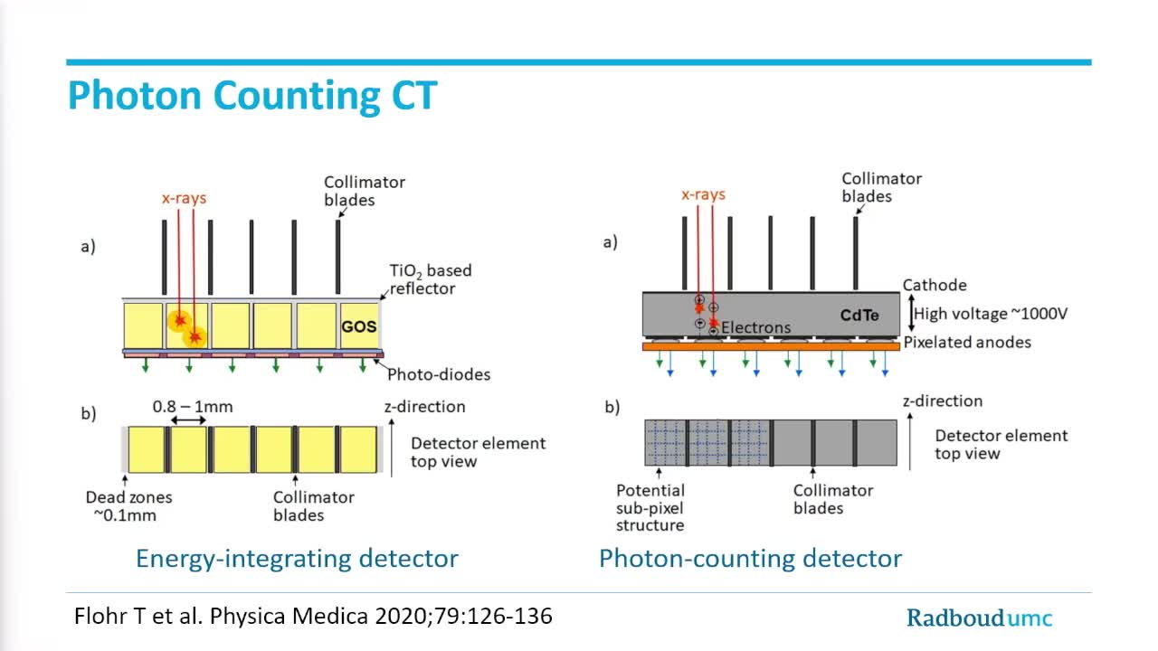
Image credit: ECR Live Stream
Detection: When X-ray photons pass through the patient's body, they interact with the detector material in the scanner. In photon counting detectors, each photon interacts with a semiconductor material, such as silicon or cadmium telluride.
Conversion: When an X-ray photon interacts with the semiconductor material, it produces a burst of electrical charge, known as electron-hole pairs. This charge is proportional to the energy of the incoming photon.
Counting: The electrical charge generated by each interaction is measured and converted into a digital signal. Each detected photon is counted individually, allowing for precise measurement of the number of X-ray photons that pass through the patient's body.
Energy Discrimination: Photon counting detectors can also discriminate between X-ray photons of different energy levels. This ability is crucial for spectral imaging applications, where information about the energy distribution of X-ray photons can provide additional diagnostic information.
Image Reconstruction: The collected data, including the number and energy of detected photons, are used to reconstruct cross-sectional images of the patient's anatomy. Advanced algorithms are employed to process the raw data and generate high-resolution images with excellent contrast and minimal noise.
By using thresholds, clinicians can remove electronic noise and focus only on the things that blip out of the noise. The limit is around 25 keV, vastly below what’s used in clinical practice. Then bins, 5 in Canon Medical’s prototype, are installed at various energy level and collect and count the photons and their energy charge. By just looking at the charge, PCCT technology makes it possible to subdivide the detector areas, thus greatly enhancing the resolution. Using 5 bins allows to optimise the images, but the amount of data is gigantic.
Nevertheless, the image quality gain potential is huge with Super-High Resolution mode, as displayed in these examples.
Cortical bone
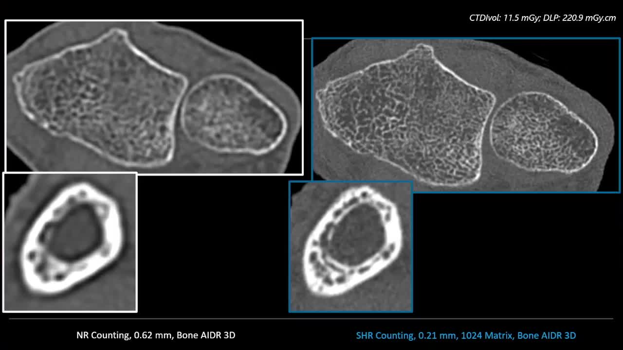
Image credit: ECR Live Stream
Global illumination rendering of fingers and elbow
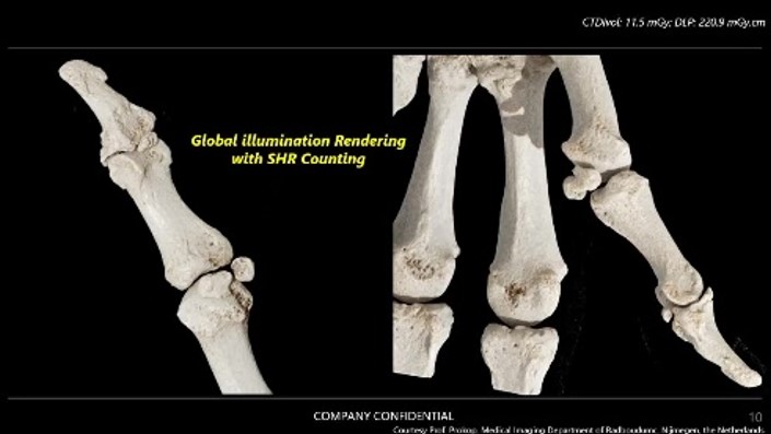
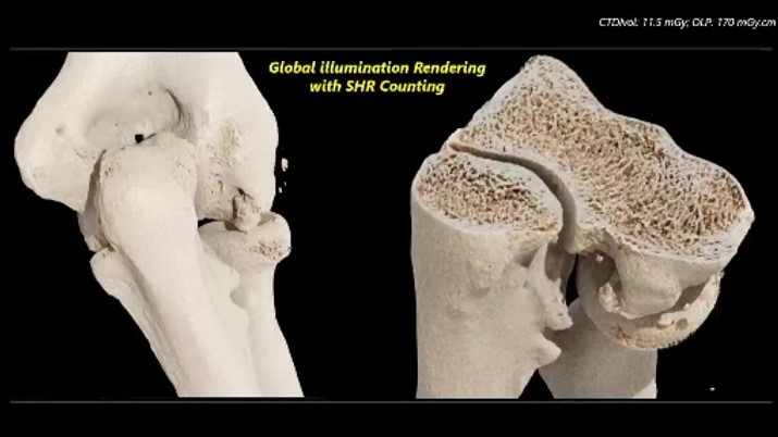
Images credit: ECR Live Stream
Bronchial cancer metastases
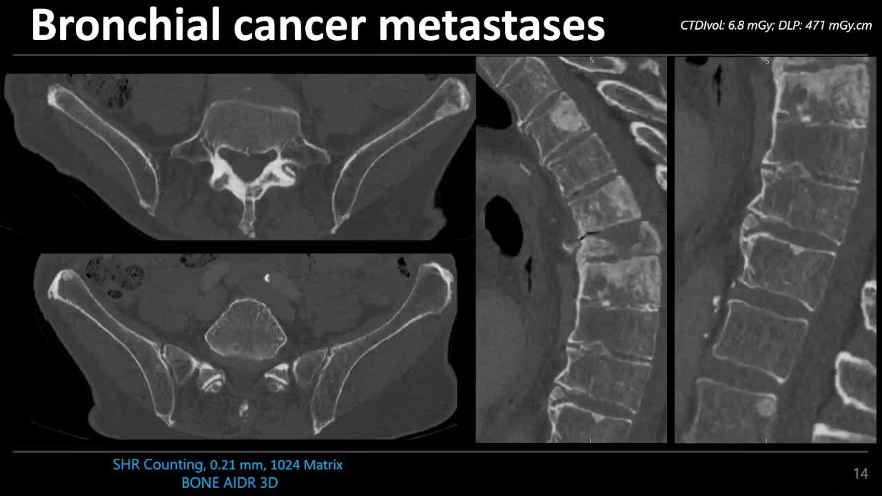
Image credit: ECR Live Stream
Inner ear
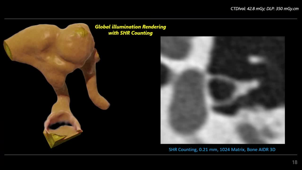
Image credit: ECR Live Stream
Chest
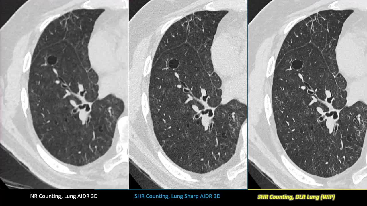
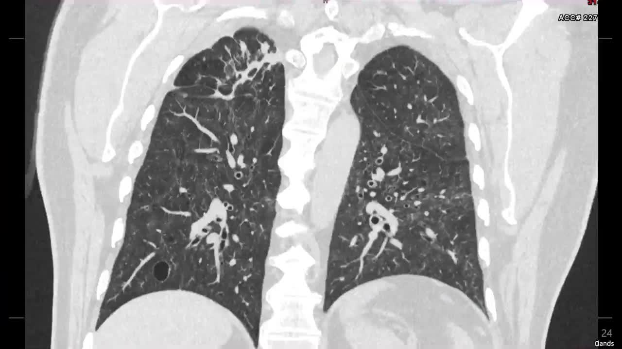

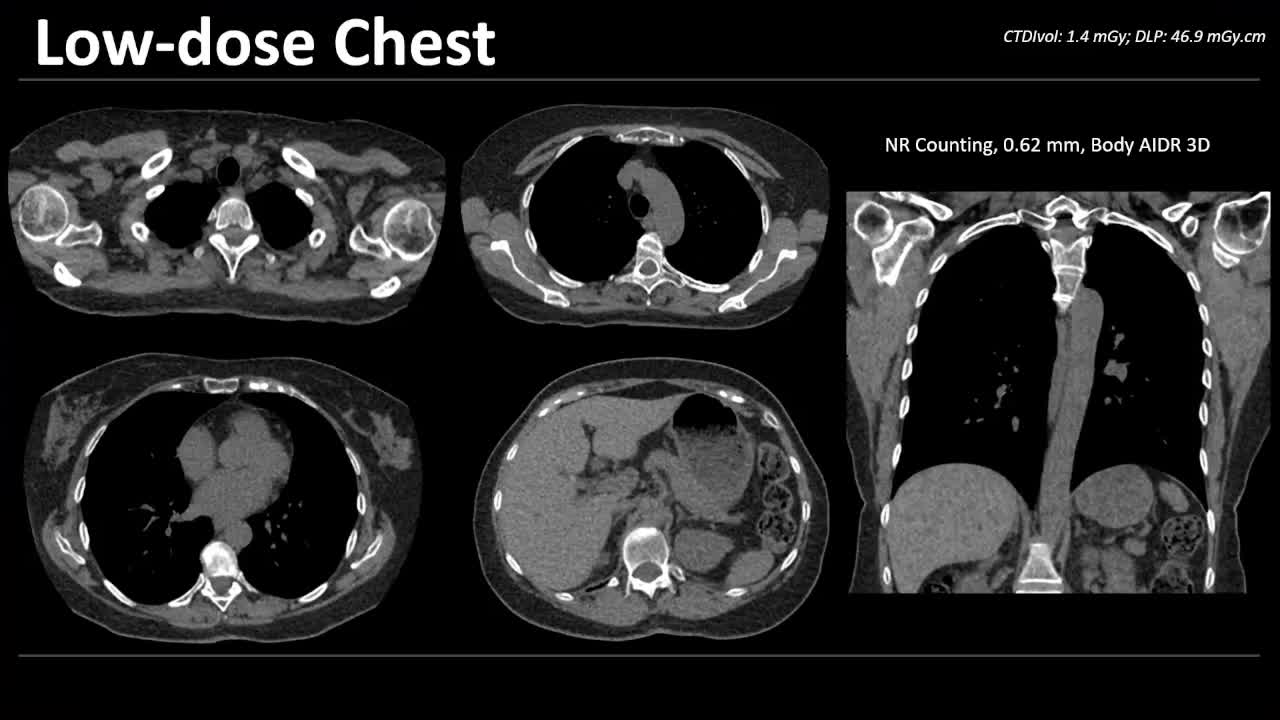
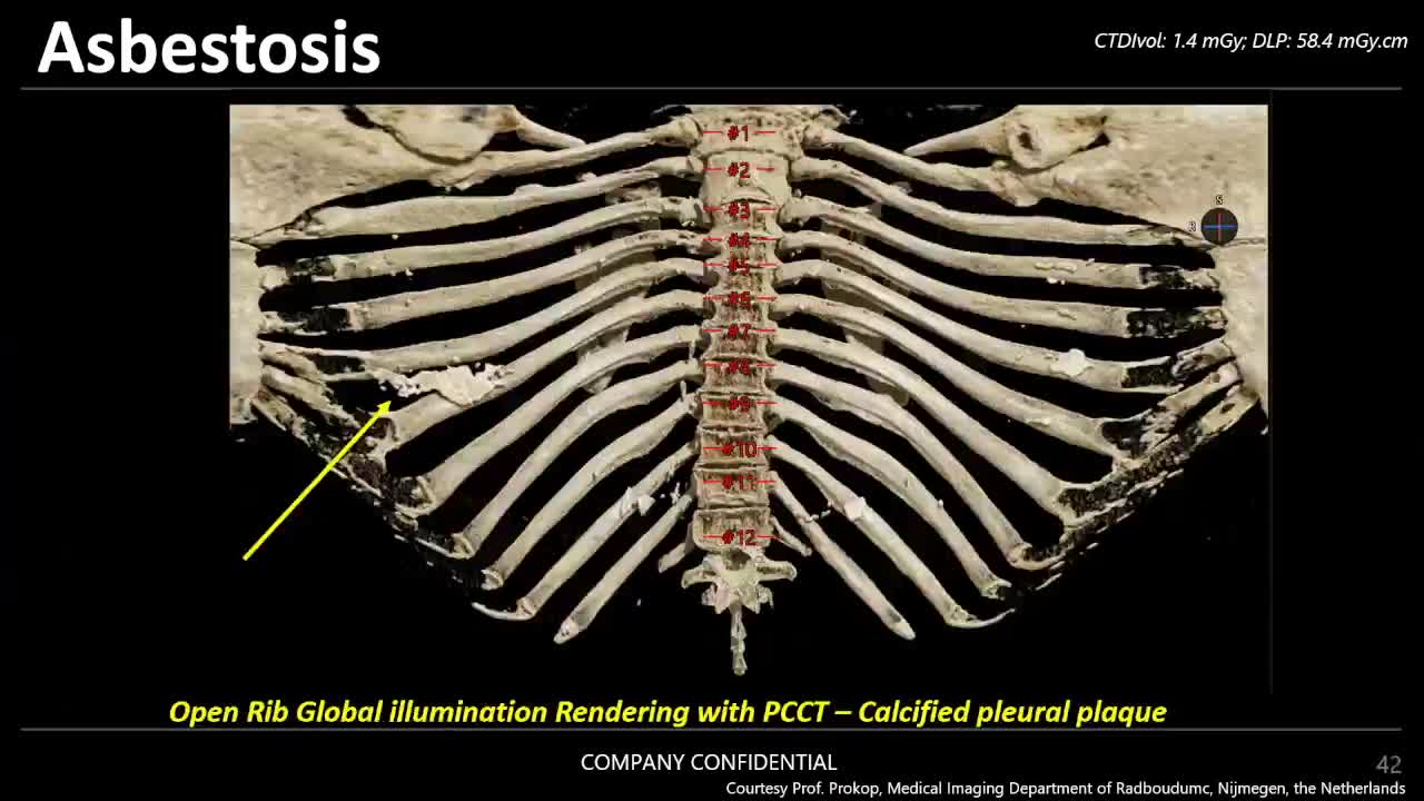
Images credit: ECR Live Stream
Abdomen
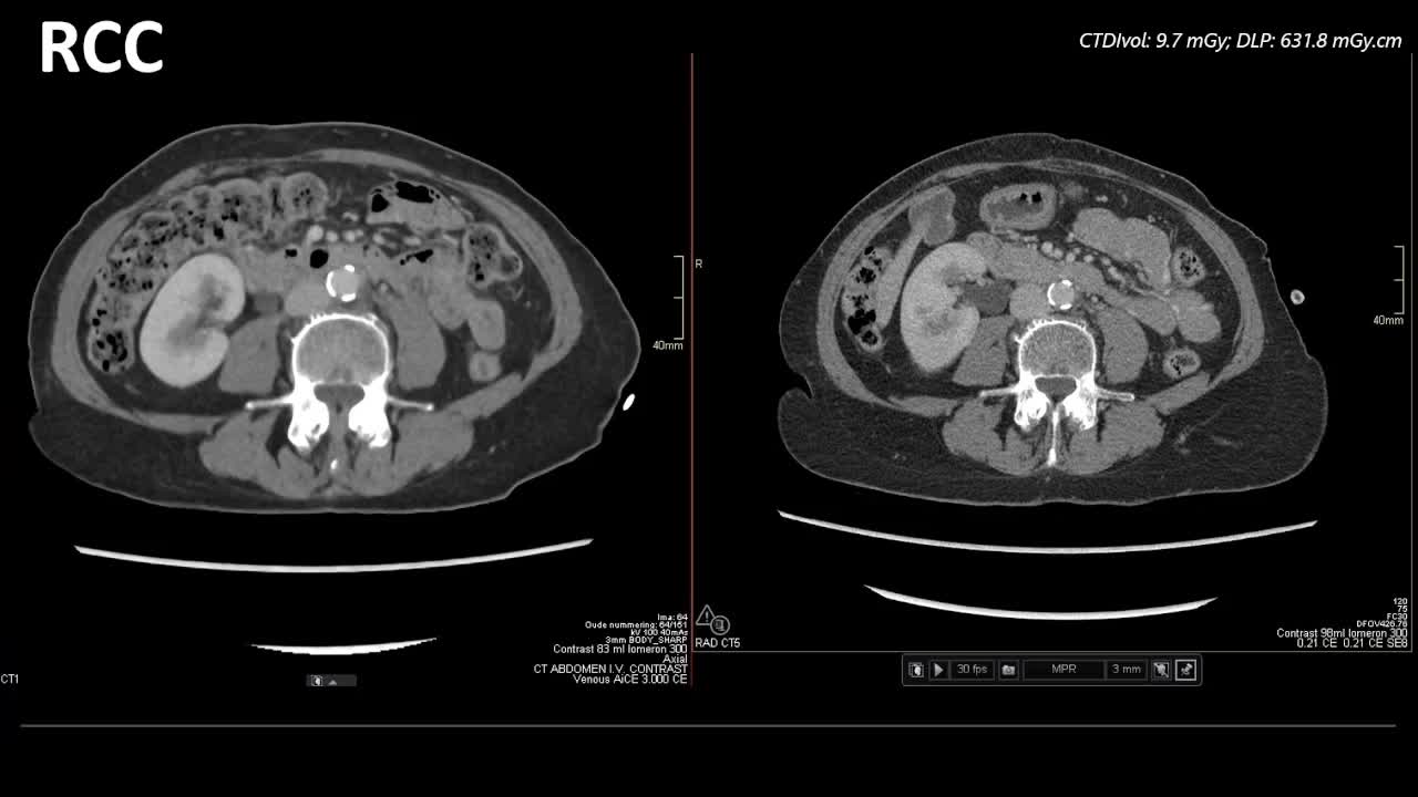
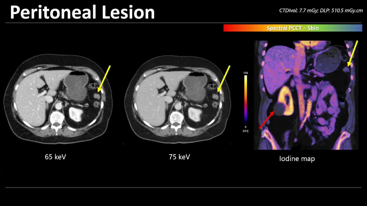
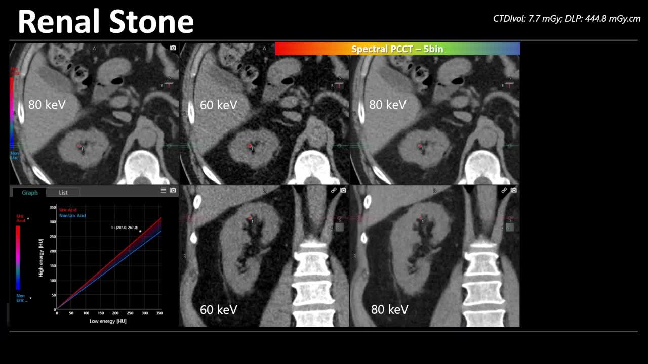
Images credit: ECR Live Stream
Through the early implementation of Canon PCCT, which brings together various technologies that Canon Medical has cultivated over the years, Canon Medical will contribute to the further development of diagnostic imaging technology and the improvement of healthcare. With Canon Medical’s innovations and technology leadership, Canon Medical will aim for the No. 1 share in the global CT market.
Cover Image Credit: Canon Medical Systems Europe
Other Image Credit: ECR Live Stream


- What Does it Mean for Radiology?")


