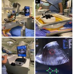
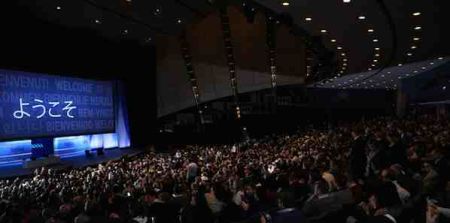
Regina Hooley, MD, Associate Professor of Diagnostic Radiology at Yale University School of Medicine was the keynote speaker yesterday on the state of the art of breast ultrasound at the Annual Scientific Meeting of the Radiological Society of North America (RSNA) in Chicago. The US modality has increasing evidence regarding its effectiveness in breast imaging.
Mammography is the gold standard, but is an imperfect test. Sensitivity can be high as 98% in fatty breasts, but as low as 30-55% in dense breasts. With increased breast density comes increased cancer risk, interval cancers and worse prognosis for clinically detected cancers.
In the United States 19 states now have mandatory breast density inform legislation, while advocates are pushing for a nationwide mandate, either through legislation (a Breast Density and Mammography Reporting Act has been introduced every year since 2011) or regulation (a decision to amend the Mammography Quality Standards Act to amend the patient letter is pending). Hooley noted that there is no consistency across states that have enacted laws. There is inconsistent mention of density as a risk factor, variable notification (all breast patients vs only those with dense breasts and varying specificity about supplemental screening (US and MRI are mentions vs “more screening” vs no mention.)
Hooley suggested there will be increased demand for screening breast US. It is complementary to mammography, performs well in dense breasts, convenient, has a low positive predictive value (PPV), but high false positives, with increased healthcare costs due to false positives. Most cancers detected on handheld screening breast US are invasive, < 1cm and node negative. The rationale is that finding mammographically occult cancers at an earlier stage and smaller size will improve overall mortality, thus allowing breast conserving therapy and less harmful treatments. There is no proven long term mortality benefit to women, as no RCT has ever been completed, however.
A recent study by Bae et al. (2014) found 335 cancers out of 116,683 screening ultrasound exams, a cancer detection rate of 3.4 per thousand. 19% (63) were found retrospectively on mammography, so were interpretive errors. However, 81% (272) had no abnormal mammographic findings. Of these, 9 cancers were in areas not included on the mammogram. Hooley noted that use of US is common in Asia, as although prevalence of breast cancer is lower in Asian woman, the incidence of high breast density is greater.
Who Should Perform Breast Ultrasound?
Technologists can perform handheld screening ultrasound, but should have experience in diagnostic/targeted US. Techs should have breast ARDMS or ARRT certification, although this is not specific to screening. Japan has a formal two day training course that includes interpretation of still images, videos and real-time screening.
Disadvantages
The disadvantages of handheld screening US include a technologist detection rate slightly lower than exams performed by a physician. Hooley explained that there is an issue of performance fatigue, as only 3-4 cancers are found for every 100000 scans. Handheld screening is operator dependent. There is also decreased reproducibility.
The disadvantages of automated screening breast US are that some units only perform whole breast US, so patients have to move to another room for different types of scan. In addition, automated breast US may not scan the entire breast. It is time-consuming to review. Patients have to be recalled for a second exam.
There are two types of equipment - either a US unit with 15 cm wide transducer or a computer-guided mechnical arm fixed to a standard US unit.
Hooley cited a recent study by Brem et al. published in Radiology. 15,318 women received a screening mammogram plus automated breast US (ABUS) 223 cancers were found, with nearly 30% (112) only detected with ABUS, giving a cancer detection rate (CDR) of 2.0 per thousand. 93% of these were invasive, the average size was 13mm, 78% were node negative. The recall rate due to US was 13.4%.
Should Screening US be Performed at the Same Time as the Mammogram?
Hooley agreed that on the same day is efficient for patients, as scheduling two appointments may be difficult. High risk anxious patients may desire imaging alternating at six monthly intervals, so they are seen more frequently.
Tohono et al.’s study in Japan concluded that it was possible to reduce the mammography recall rate when combining mammography with screening US. If a mass was seen on a mammogram, the woman waited for US. 11,753 women were screened. Of these, 974 women had mammography and US performed on the same day. The mammography recall rate/ CDR were as follows:
- Mammo only: 4.9%/0.22
- Mammo + uS: 2.6%/0.31
Benign Findings
Hooley noted that cysts were seen in nearly half of participants in the ACRIN trial; 14% of participants had complicated cysts. There is no need to image all these findings and 12-month follow up is adequate for most. The exceptions are solitary or new cystic appearing masses in BRCA+ women, high risk women or women with a history of lymphoma or melanoma.
The ACRIN 6666 trial found low malignancy rate (0.8%) (Barr et al. 2013) in BI-RADS category lesions. Multiple bilateral circumscribed masses were three times more common with US compared to mammography (Berg et al. 2013 - http://www.ncbi.nlm.nih.gov/pubmed/23616634), with no malignancies at two years. 12-month diagnostic follow up may be appropriate in these cases.
Shearwave Elastography (SWE)
SWE may be useful to add value for the evaluation of breast masses detected with screening, thus decreasing the need for biopsy of BI-RADS 4a lesions, thereby increasing PPV. A study in Radiology by Lee et al. in 2014 found increased specificity in 195 BI-RADS 4a lesions. They found that the following criterion allowed reclassification as BI-RADS 3: dark blue colour or less than or equal to 30 KPa meant no cancers. This improved specificity from 9% to 59-57%, with no loss in sensitivity.
Tomosynthesis
Hooley put the question - with the availability of digital breast tomosynthesis (DBT) is there a need for screening US. She concluded that
with either tomosynthesis or screening US the cancer detection rate will increase compared to full-field digital mammography alone. Both modalities will detect mostly small invasive cancers. Ultrasound is primarily a second exam, for dense breasts, with a low positive prediction value. DBT with 2D mammography is only one exam as far as the patient is concerned and can handle all breast densities. It has fewer recalls and a higher positive predictive value. However, the cost-effectiveness and cancer detection rate of supplemental screening US may decrease.
Hooley concluded that more research is needed on breast US to improve specificity thus increasing positive predictive value and decreasing recalls. In addition work is needed to determine its cost-effectiveness in comparison to new screening tools, such as tomosynthesis and fast MR.
Claire Pillar
Managing Editor, HealthManagement
Latest Articles
Regina Hooley, MD, Associate Professor of Diagnostic Radiology at Yale University School of Medicine was the keynote speaker yesterday on the state of the...


