


New 3D printed heart technology is to be showcased at EuroEcho-Imaging 2014, the official annual meeting of the European Association of Cardiovascular Imaging (EACVI). EACVI is also a registered branch of the European Society of Cardiology (ESC). The congress is scheduled to take place between 3-6 December in Vienna, Austria at the Reed Messe Wein GmbH Congress Center.
The latest research and clinical findings on echocardiography and other cardiovascular imaging techniques will be presented and discussed at the conference, one of the leading congresses for clinicians and scientists. The most up-to-date findings in the field are expected to be presented over four days of scientific sessions. In addition, new frontiers in cardiovascular imaging will be explored with presentations on three dimensional imaging, a central theme of the congress.
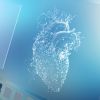
3D Printed Heart Model
Experts will also reveal technology that uses imaging data to print a 3D model of the heart. Participants will have first-hand access to experts who have experience of this cutting edge technique. According to Professor Patrizio Lancelloti, EACVI President, "The heart is a 3D structure that we traditionally analysed using 2D imaging including echocardiography, cardiac magnetic resonance imaging (CMRI) or cardiac computed tomography (CT). But with the advent of 3D imaging, now we can clearly evaluate the structure of the heart in different planes."
Professor Lancelloti also points out that the ability to print a 3D model of the heart is a major advancement in this field and can go a long way in helping clinicians perform surgical and percutaneous interventions on the mitral or aortic valve, choosing the perfect size of device and planning the procedure according to the patient's unique anatomy. He believes that the ability to create a 3D computational model of the heart will not only assist with interventions, but will also improve the understanding of the heart's physiology by providing greater insight into the interactions between the valves and the ventricles, the ventricles and the aorta, and the valves and the left atrium.
Imaging in Acute Care
The second major theme of the congress is imaging in acute care, including key topics such as stress imaging to rule out myocardial ischaemia. Today, echocardiography is the primary method that is used for diagnosing patients with unstable cardiovascular disease in the acute setting. During the congress, cardiac oncology will be the subject of a detailed session in which novel findings on how to detect and prevent the effects of cancer drugs on the heart will be presented. Recommendations for monitoring cardiotoxicity after radiotherapy and chemotherapy will also be discussed.
Prof Lancellotti said: "More cancer patients are surviving cancer and living longer but then developing cardiac disease. Imaging plays a major role in the diagnosis and follow up of these patients. The ESC/EACVI's Cardiac Oncology Toxicity Registry was launched in September to collect data on practices for identifying and treating cardiotoxicity of breast cancer drugs and progress will be reported at EuroEcho-Imaging."
The congress will also include the release of ground-breaking research abstracts. Oral abstracts will be presented in the Agoara area. The Roelandt Young Investigator Awards will also be launched to honour the late Jos Roelandt, the founding editor of the society's journal. Joint sessions with several sister societies across the globe will also be held.
Source: European Society of Cardiology
Image Credit: InternetMedicine.com
Latest Articles
New 3D printed heart technology is to be showcased at EuroEcho-Imaging 2014, the official annual meeting of the European Association of Cardiovascular Imag...