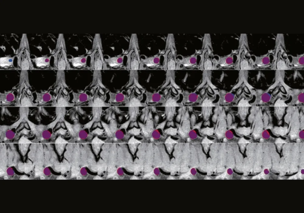
In the pursuit of automating the detection and segmentation of intracranial aneurysms (IAs), a recent study published in European Radiology aimed to address the challenge of automating the segmentation and detection of IAs using deep learning techniques, specifically focusing on magnetic resonance T1 images. Through a retrospective diagnostic investigation involving 159 IAs from 136 patients who had undergone T1 imaging, the researchers designed and evaluated a deep learning framework tailored for this purpose. Data from 127 cases were utilised for training and validation, while an additional 32 cases were set aside for assessing the algorithm's accuracy and consistency. Leveraging convolutional neural networks (CNNs), three distinct models were developed and assembled to perform the segmentation and detection tasks. To evaluate the model's performance, various metrics were computed across different levels, including at the voxel, IAs, and patient levels, providing a comprehensive assessment against ground truth data.
Encouraging Results: Robustness and Clinical Relevance
The results of the study are highly encouraging, demonstrating the promising performance of the deep learning framework. Notable metrics such as an overall Dice score of 0.802, voxel-level sensitivity of 0.874, specificity of 0.9998, balanced accuracy of 0.937, and an F1 score of 0.802 speak to the robustness of the approach. A threshold of 0.7 for coincidence between predicted and ground truth aneurysms was used to identify true positives. Furthermore, the model exhibited a sensitivity of 90.63% for detection, accompanied by a low false positive rate of 0.58 per case. Importantly, the volumes of IAs segmented by the model exhibited strong agreement and consistency with volumes labelled by expert clinicians, further bolstering confidence in the framework's potential clinical applicability.
Advancing Medical Imaging Through Tailored Segmentation
Key insights from the study include the novel contribution of a segmentation method tailored specifically for clinical T1 images, filling a previously identified gap in existing literature. Additionally, the universality of T1-based segmentation and detection methods was emphasised, offering potential advantages in scenarios where angiography images are unavailable. Finally, the robustness and clinical applicability of the developed deep learning framework position it as a promising tool for enhancing the detection and management of intracranial aneurysms in real-world clinical practice.
Future Directions: Beyond Traditional Imaging Modalities
The deep learning framework not only demonstrated feasibility and robustness in the segmentation and detection of IAs using routine T1 sequences, but also showcased its potential to outperform existing methods reliant on digital subtraction angiography (DSA), computed tomography angiography (CTA), and magnetic resonance angiography (MRA). Importantly, the framework's ability to leverage T1 images for effective identification and segmentation of intracranial aneurysms presents a beacon of hope for early detection and intervention in clinical settings, potentially revolutionising the field of medical imaging.
Source: European Radiology
Image Credit: ESR


