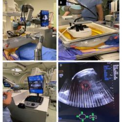
A study published in Radiology aimed to establish correlations between quantitative ultrasound (US) measures (like shear-wave speed and acoustic attenuation) and MRI measures (like liver stiffness and PDFF) in children, adolescents, and young adults with known or suspected chronic liver disease. The goal was also to determine the predictive ability of US measures in detecting abnormal liver stiffening and steatosis, using MRI as the reference standard.
The Role of Quantitative Imaging in Liver Disease Assessment
Paediatric chronic liver disease can be caused by various conditions, with non-alcoholic fatty liver disease being the most prevalent in children. The progression of most chronic liver diseases leads to fibrosis, cirrhosis, and eventually liver failure. Early stages of the disease can present with steatosis, congestion, and inflammation. While liver biopsy is the gold standard for diagnosis and severity assessment, imaging techniques like US shear-wave elastography (SWE) and MR elastography are increasingly used to detect liver stiffening indicative of fibrosis and differentiate between fibrosis and simple steatosis. There is extensive research supporting SWE and MR elastography as quantitative measures for liver stiffening due to fibrosis, as well as MRI liver proton density fat fraction (PDFF) for quantifying hepatic steatosis. US manufacturers are exploring additional quantitative techniques to identify diffuse liver disease changes, but data on their diagnostic performance and relation to MRI measures are limited.
Study Participants and Demographics
Out of 70 patients contacted for the study, 45 agreed to participate. However, one female participant was excluded due to discomfort during MRI, leaving 44 participants (23 males and 21 females) for evaluation. Technical issues prevented the collection of NLV and hepatorenal index data for two participants, and GRE MR elastography data was unanalysable for one participant due to high liver iron content. Additionally, eight measurements (one attenuation and seven individual hepatorenal index measurements) were excluded due to technical inadequacy. The most common liver diseases among participants were Fontan-associated liver disease (16%), known fatty liver disease (14%), and autoimmune liver disease (14%). The average age of participants was 16 years with a BMI of 21.9 kg/m2. The mean liver shear stiffness measured by GRE MR elastography was 3.3 kPa, and the mean liver proton density fat fraction (PDFF) was 5.6%. Liver shear stiffness was found to be higher in males (3.8 kPa) compared to females (2.7 kPa), and liver PDFF was also higher in males (5.8%) than females (5.3%).
Key Findings on Ultrasound and MRI Correlations
Quantitative MRI and US are increasingly important in evaluating liver disease, but there's limited data comparing these modalities, especially concerning newer US techniques. This study aimed to assess the diagnostic performance of quantitative US in children with chronic liver disease using MRI as a reference. In the study, US-assessed median liver shear-wave speed (SWS) and shear-wave dispersion correlated positively and moderately to highly with liver shear stiffness measured by MR elastography. SWS was identified as the sole predictor of abnormal MRI liver shear stiffness (>2.8 kPa), with an AUC of 0.95. Median US acoustic attenuation moderately correlated with MRI proton density fat fraction (PDFF), and was an independent predictor for abnormal liver fat (>5%), with an AUC of 0.75.
Significance of Shear-Wave Dispersion and Attenuation Coefficient
Comparing with previous studies, the correlation between US liver SWS and MRI liver shear stiffness in this study was higher. The variations could be due to technique differences, US manufacturer differences, or specific BMI inclusion criteria. The study also found a high correlation between US SWS and shear-wave dispersion. However, more research is needed to determine the utility of dispersion as a complementary imaging marker. The positive association between US attenuation coefficient and MRI PDFF aligns with existing literature. The study acknowledges limitations such as a small sample size, lack of liver biopsy as a reference standard, and recruitment emphasising liver shear stiffness over hepatic steatosis range.
The study demonstrated a moderate to high correlation between US liver SWS and MR elastography-derived stiffness, and a moderate correlation between US attenuation coefficient and liver MRI PDFF in young individuals with a BMI less than 35 kg/m2 and chronic liver diseases. Validation and further research across different US system manufacturers are needed.
Source & Image Credit: RSNA Radiology


