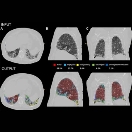
A recent study published in Radiology sheds light on the complex landscape of pulmonary hypertension (PH), particularly in distinguishing between pulmonary arterial hypertension (PAH) and PH associated with lung disease (PH-LD). The severity of lung disease plays a crucial role in this differentiation, with significant implications for treatment eligibility and prognosis.
Advancing Beyond Radiological Scoring
Traditional methods of assessing lung disease severity through radiological scoring have limitations, including poor reproducibility among radiologists. To address this challenge, researchers investigated the potential of artificial intelligence (AI) to quantify lung fibrosis using chest CT imaging. The study, led by Dwivedi et al., aimed to evaluate the prognostic value of AI-quantified fibrosis and its combination with radiological scoring in predicting survival outcomes. The study analysed data from the ASPIRE registry, which includes patients diagnosed with incidental PAH and PH-LD. Eligible patients underwent baseline CT imaging, and exclusion criteria ensured a comprehensive representation of lung disease severity and PH aetiology. The AI model, trained on a deep learning architecture, accurately quantified fibrosis percentage and other parenchymal patterns from CT scans.
Enhanced Prognostication with AI-Quantified Fibrosis
Results revealed that AI-quantified fibrosis was independently associated with increased mortality risk, even after accounting for demographic and hemodynamic factors. Importantly, the combination of AI-quantified fibrosis and radiological scoring significantly enhanced prognostic accuracy compared to radiological scoring alone. The AI model's ability to detect subtle parenchymal changes provided additional predictive value, especially in patients radiologically scored as having no fibrosis. The study's findings hold promise for improving patient phenotyping and treatment stratification in PH. By better characterising lung disease severity, clinicians can identify individuals likely to benefit from targeted therapies and monitor disease progression more effectively. The AI model, designed as an adjunct to radiological reporting, offers a transparent and interpretable tool for integrating quantitative lung assessment into routine clinical practice.
Challenges and Future Directions in AI-Driven Lung Disease Assessment
Despite the study's strengths, including its large dataset and external validation, several limitations should be acknowledged. Retrospective data analysis and variations in CT imaging protocols may introduce bias. Additionally, while the AI model demonstrated robust performance, its generalizability to different settings requires further validation. In conclusion, the study underscores the potential of AI-driven quantitative analysis to enhance prognostication in PH patients. Future research endeavours should focus on prospective multicentre studies to validate the utility of AI models in refining patient phenotyping and optimising treatment strategies in PH and lung disease management.
Source & Image Credit: RSNA- Radiology


