Radiologist Dr Georgia Spear of NorthShore Medical Group
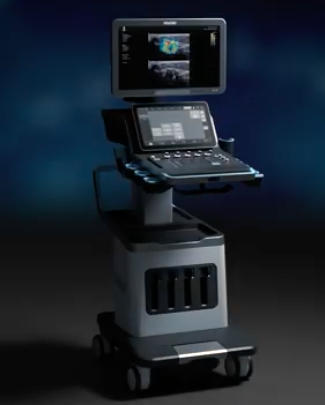
The Hologic’s SuperSonic™ MACH™ 40 Ultrasound System brings radiologists a new generation of ultrasound technology.
At times, mammographic findings turn out very subtle. Ultrasounds enhance the detection of these findings, thus improving the accuracy of diagnosis.
This new technology provides images with reduced speckles, and assists clinicians to see lesions that are not normally seen before, including in patients with dense breast tissue. As a result of the image quality and resolution, the confidence level in diagnosis has vastly improved.
This technology helps clinicians on multiple levels. For example, a new radiologist might find it beneficial to use the technology’s ShearWave elastography during lesion characterisation. This can help inform the clinician about the tissue stiffness, enabling them to accurately estimate the tumour size. This is particularly useful for deciding whether a six-month follow-up is necessary or whether to perform a biopsy on a particular patient.
For long-time practicing clinicians, ShearWave elastography can help them to correlate the lesion characterisation with the biopsy results. In clinical practice, this assists them with arriving at a diagnosis faster and getting patients the immediate treatment they require.
This new technology eliminates the most common concerns a radiologist would normally have with old technology. Firstly, it can be heavily relied on during procedures and diagnostic interpretations. For example, during localisation procedures, one looks at very small lesions that have a biopsy marker or focuses on axillary lymph nodes where biopsy markers are often placed. It can be very difficult to detect and gain a visual of the biopsy markers, but with this new technology, radiologists can readily see them. One can then target the lesions more efficiently, quickly and effectively.
Secondly, the practical benefits hugely improve clinician experience. The longevity of this unit means that it can work reliably throughout the day, and clinicians no longer have to feel concerned the system will shut down during their practice.
It is quick and customisable. The monitor can move around, which allows clinicians to place it in the perfect location to use the best angle during examinations. This has had a positive impact as most technologists encounter practical discomfort problems, including hunching and constant attempts to move the screen around. Now the system can be brought up to the level the clinicians need, so they are no longer straining.
The overall goal in breast imagery practice is to detect breast cancers early when it is manageable and treatable. The Hologic SuperSonic™ MACH™ 40 Ultrasound System is easy to navigate and incredibly efficient for arriving at the diagnosis faster.
Source: Hologic