


In a first, researchers have used CT imaging and 3D printing in the surgical planning for separation of conjoined twins, according to a study presented on Wednesday, 2 December, at the annual meeting of the Radiological Society of North America (RSNA). The 3D models proved to be an excellent source of information, as there were no major discrepancies between the models and the twins' actual anatomy.
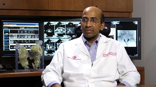
"The surgeons found the landmarks for the liver, hearts and pelvic organs just as we had described," said lead author Rajesh Krishnamurthy, MD, chief of radiology research and cardiac imaging at Texas Children's Hospital in Houston. "The concordance was almost perfect."
Conjoined twins, or twins whose bodies are connected, occur once every 200,000 live births and their survival rates are low. Separating them through surgery is extremely difficult because they often share organs and blood vessels.
To prepare for surgery to separate the conjoined twins — connected from the chest all the way down to the pelvis — Dr. Krishnamurthy and colleagues performed volumetric CT imaging with a 320-detector scanner. The team administered intravenous contrast separately to both twins to enhance views of vital structures and help plan how to separate them to ensure survival of both children. They used a technique known as target mode prospective EKG gating to freeze the motion of the hearts on the images and get a more detailed view of the cardiovascular anatomy, while keeping the radiation exposure low.
CT scans showed that the babies' hearts were in the same cavity but were not fused. "Also, we detected a plane of separation of the liver that the surgeons would be able to use," Dr. Krishnamurthy revealed.
The official separation of the 10-month-old twins took place approximately 18 hours into the 26-hour surgery. The operation was performed by a team of more than 26 clinicians, including 12 surgeons, six anaesthesiologists and eight surgical nurses. The twins have returned home and are both doing well.
Dr. Krishnamurthy expects the combination of volumetric CT, 3D modelling, and 3D printing to become a standard part of preparation for surgical separation of conjoined twins, although barriers remain to its adoption.
"The 3D printing technology has advanced quite a bit, and the costs are declining. What's limiting it is a lack of reimbursement for these services," he noted. "The procedure is not currently recognised by insurance companies, so right now hospitals are supporting the costs."
Iphigenia Papaioanou
Editorial Project Director, HealthManagement.org
Source and image credit: RSNA
"The surgeons found the landmarks for the liver, hearts and pelvic organs just as we had described," said lead author Rajesh Krishnamurthy, MD, chief of radiology research and cardiac imaging at Texas Children's Hospital in Houston. "The concordance was almost perfect."
Conjoined twins, or twins whose bodies are connected, occur once every 200,000 live births and their survival rates are low. Separating them through surgery is extremely difficult because they often share organs and blood vessels.
To prepare for surgery to separate the conjoined twins — connected from the chest all the way down to the pelvis — Dr. Krishnamurthy and colleagues performed volumetric CT imaging with a 320-detector scanner. The team administered intravenous contrast separately to both twins to enhance views of vital structures and help plan how to separate them to ensure survival of both children. They used a technique known as target mode prospective EKG gating to freeze the motion of the hearts on the images and get a more detailed view of the cardiovascular anatomy, while keeping the radiation exposure low.
CT scans showed that the babies' hearts were in the same cavity but were not fused. "Also, we detected a plane of separation of the liver that the surgeons would be able to use," Dr. Krishnamurthy revealed.
The official separation of the 10-month-old twins took place approximately 18 hours into the 26-hour surgery. The operation was performed by a team of more than 26 clinicians, including 12 surgeons, six anaesthesiologists and eight surgical nurses. The twins have returned home and are both doing well.
Dr. Krishnamurthy expects the combination of volumetric CT, 3D modelling, and 3D printing to become a standard part of preparation for surgical separation of conjoined twins, although barriers remain to its adoption.
"The 3D printing technology has advanced quite a bit, and the costs are declining. What's limiting it is a lack of reimbursement for these services," he noted. "The procedure is not currently recognised by insurance companies, so right now hospitals are supporting the costs."
Figure 1. Dr. Krishnamurthy with his 3D model.
Iphigenia Papaioanou
Editorial Project Director, HealthManagement.org
Source and image credit: RSNA
Latest Articles
healthmanagement, #RSNA15, RSNA 2015, CT scans, 3D printing, surgery, reimbursement
In a first, researchers have used CT imaging and 3D printing in the surgical planning for separation of conjoined twins, according to a study presented on Tuesday, 1 December, at the annual meeting of the Radiological Society of North America (RSNA).


