HealthManagement, Volume 21 - Issue 2, 2021
 PRINT OPTIMISED
Key Points
- SmartPositioning augments a live camera stream by projecting the image area and the AEC x-ray feedback sensor positions onto the patient’s body.
- SmartRotate uses deep learning to auto-rotate images to their standard orientation based on the image content.
- SmartAlign uses advanced sensing to give live feedback on the accuracy of the tube-to-panel alignment during bedside or out-of-bucky exams.
- SmartDose uses 3D machine vision to determine the thickness of the patient, and then tailors the exposure parameters.
- Agfa’s SmartXR portfolio, comprising SmartPositioning, SmartRotate, SmartAlign and SmartDose, is underpinned by artificial intelligence (AI) solutions that help radiology departments meet their workflow challenges by improving operational efficiency and clinical consistency.
Introduction
X-rays are the single most performed imaging diagnostic: every year more than two million radiographic exams are carried out on Agfa equipment around the world. Radiology and radiography workloads are continuously growing, while the move to evidence-based medicine is simultaneously increasing the pressure on healthcare budgets, efficiency and quality of care standards.
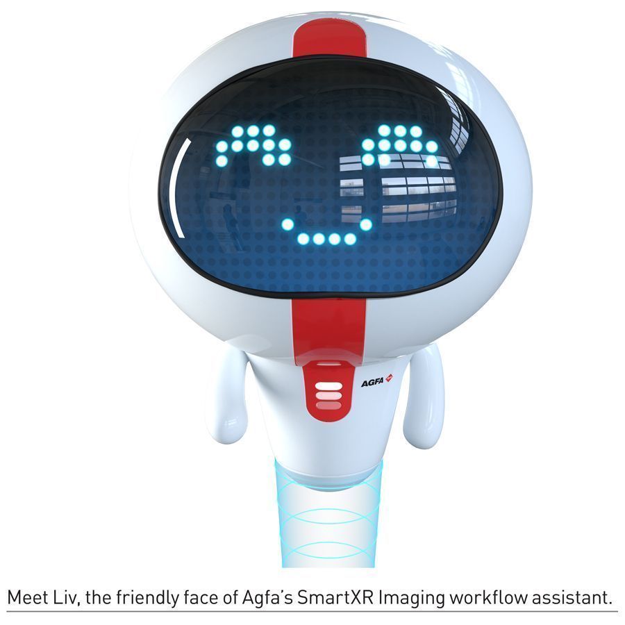
With its SmartXR portfolio, Agfa has directed its development of artificial intelligence (AI) solutions towards helping radiology departments meet their workflow challenges. These AI upgrades for digital radiography focus on supporting operational efficiency and clinical consistency. We interviewed Gert Merckx, Product Manager Radiology Solutions, and Jeroen Cant, Research Team Lead, from Agfa, to find out more.
SmartXR at a glance
• Smart Positioning augments a live camera stream of the patient by projecting the image area and the AEC x-ray feedback sensor positions onto the patient’s body. This helps the technician speed up positioning, while making it more accurate and consistent to reduce retakes.
• Smart Rotate uses deep learning to auto-rotate images to their standard orientation based on the image content. Fewer post-processing actions are needed, while image presentation on both workstation and PACS is more consistent.
• Smart Align uses advanced sensing to give live feedback on the accuracy of the tube-to-panel alignment during bedside or out-of-bucky exams. Alignment is faster and more accurate, with fewer retakes due to grid cut-off and more consistent projections.
• Smart Dose uses 3D machine vision to determine the thickness of the patient, and then tailors the exposure parameters. This helps the technician to speed up the configuration of exposure settings, reduce retakes, improve image consistency and ensure the ideal dose for the patient.
You have described SmartXR as an “intelligent radiography workflow assistant”. Why did Agfa choose to focus its artificial intelligence efforts on workflow, rather than diagnostic AI, for example?
Gert Merckx: Artificial intelligence has opened up a lot of possibilities for radiology. One area that has received a lot of attention is diagnosis, for example, to detect tuberculosis or pneumothorax. This is a very important functionality; there are a number of vendors working on it.
But, we chose to take a different approach, and to use our strengths and 120-year legacy in radiography. Since the era of film, optimising workflow and reducing waste have always been a key focus for our solutions and services. So, it made sense for us to develop tools and functionalities that address the most pressing challenges facing radiology departments.

One of these is a shortage of skilled technicians, combined with the fact that students these days receive fairly limited training on x-ray radiography. This raises problems that impact quality and efficiency, including avoidable errors, high exam variability and more. For the technicians, the situation can also create a lot of nuisance. They have to spend more time fussing with equipment, rather than focusing their attention on what really matters: the patient.
What exactly is SmartXR, and how does it help overcome the challenges facing the radiology department?
Gert Merckx: SmartXR is a portfolio of software upgrades for our direct radiography (DR) solutions. It uses the power of AI to assist the technician with very specific tasks that can be time-consuming or open to variability or errors.
We like to say that medical image acquisition is both a science and an art. SmartXR lightens the radiographer’s workload, and, at the same time, enables greater consistency in imaging, even between different technicians. So, it helps to improve the operational and clinical performance of the department, technician and radiologist.
SmartXR does this by combining hardware, such as integrated sensors and cameras, with AI-powered software, 3D machine vision, deep learning and machine intelligence. By reducing the need for retakes at different points in the technician’s workflow, SmartXR helps to eliminate waste, support quality care and enable a more efficient workflow.
Some of the technologies come from the military and gaming industries, which are quite advanced and proven. So, we could focus our minds and resources on making sure it meets today’s healthcare needs.
In the era of digital radiography, why do we have to worry about retakes?
Gert Merckx: In the classic film era, retakes had a very obvious cost: the expense of the film, time and products needed to develop the images. We created programmes to help technicians reduce the number of retakes, by optimising their imaging techniques.
But, when digital radiography was adopted, there was no longer an “out of pocket” expense for retakes. Technicians could take as many images as needed to get the right one.
However, retakes still have hidden costs: in efficiency and safety, for example. Academic studies show that hospitals can have as high a retake rate as 20%. That works out to 24 days of surplus work for an x-ray system each year. Clearly, this is an area where there is a real opportunity for improvement.
How did you select which features to include in SmartXR, and how did you incorporate the “voice of the user”?
Gert Merckx: My original role at Agfa was as an image processing researcher, working with x-ray equipment in the laboratory. I was not trained to deal with the intricacies of radiographic positioning or parameter setting. So, I made a lot of mistakes. I realised that AI offered ways to overcome these types of errors and inefficiencies. Not only for me in the laboratory, but also for technicians in the radiology department.
We started by looking at existing data to see if there was a real opportunity for AI solutions to play a role. We data-mined our workstation usage patterns, for example, to see how many retakes are caused by specific issues: positioning, alignment, etc. We also checked the academic data available. Based on this, we identified first ideas for functions.
Then we took these ideas to some of our customers directly to see what they needed and wanted. We started discussions, organised workshops to talk about their pain points and presented some possibilities to them. We confirmed that the greatest interest was in AI tools for optimising dose, improving radiographic positioning, reducing variance and enabling consistent imaging.
How did you turn those ideas and interests into practical solutions, for consistent imaging, for example?
Jeroen Cant: Improving consistency has always been a priority for Agfa: MUSICA image processing has been doing it for decades. With SmartXR, we are widening the concept of consistency to other parts of the image acquisition workflow.
The very first AI applications involved teaching an algorithm to recognise something from an image: a dog versus a cat. In healthcare, some of the first applications for radiology were to recognise specific pathologies in lung images: pneumonia, pneumothorax and others. These are known as “A to B” systems: you give the algorithm a certain number of images (“A”), and then a label comes out (“B”). It’s like a child learning to recognise different animals in their environment.
For us as well, we started with image recognition, with SmartRotate. Correct image rotation is a real pain point for radiologists when they are comparing images from different days, for example. So, SmartRotate recognises the image content: is it a hand or a lung? It then rotates the image to the standard orientation for that type of image. It is a simple idea: it enhances the consistency which the radiologist needs, while simplifying the technician’s workflow.
Studies have shown that poor x-ray positioning leads to as much as 68% of rejected images. How does SmartXR address that?
Gert Merckx: Indeed, the data led us to another key priority for SmartXR: patient positioning. Ensuring correct radiographic positioning can be quite difficult for the technician, who, for example does not know the exact angle of the x-ray panel to the tube. This is especially true for bedside imaging. But, the radiologist can better compare images taken from the same angle. Again, consistency is crucial. Our SmartAlign helps the technician ensure that the x-ray beams have a consistent angle towards the panel.
In x-ray rooms, patient movement and the position of the Automatic Exposure Control (AEC) cells pose another challenge: the patient’s body blocks the technician from seeing the AEC placement. SmartPositioning solves this by projecting the AEC cells in augmented reality (AR) onto the patient, enabling the technician to immediately see if the configuration is correct.
The camera livestream with AR overlays allows the radiographer to check the positioning from the viewpoint of the system remotely, before pressing the exposure button.
AEC and Shielding: The Role of SmartPositioning
One problem with AECs identified by the AAPM and the American College of Radiologists (ACR) and discussed in a 2020 debate is gonadal shielding. When the shielding covers an active AEC cell, the cell does not receive radiation. Thus, it never indicates that adequate exposure has been achieved. The result is overexposure, and the need for a retake. This situation has become so common that the ACR has changed their recommendations to omit the use of foetal and gonadal shielding.
SmartPositioning, however, solves this problem by clearly showing the technician that an active AEC cell is covered by the shield. The technician can then disable the cell, while still using the shielding. In contrast, an active cell that is not covered by the patient will lead to underexposure: again, requiring a retake. SmartPositioning notifies the technician that the cell is uncovered, so it may be disabled.
Dose matters, regardless of patient size: what is SmartDose, and how does it work?
Gert Merckx: At Agfa we know: dose is everyone’s business. For instance, one publication showed that a “lean” patient who receives the standard dose can receive up to six times the dose they actually require. This is just one example to illustrate why ensuring the correct exposure parameters is critical.
Back in the days of film, the technician would use callipers to measure the patient’s “thickness” and then work out an appropriate, tailored dose. This practice has largely been forgotten, and incorrect exposure parameters now account for 18% of retakes. With SmartDose, there is no more need for callipers. SmartDose gives our systems “depth vision” to estimate the patient’s thickness and then suggests the best dose to the technician.
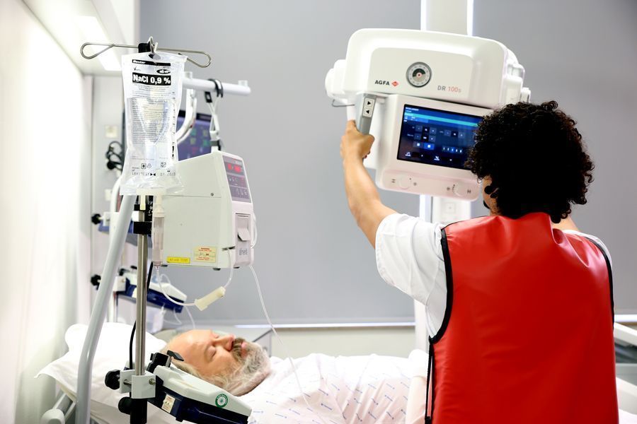
A Case in Point: SmartXR in the ICU
The global COVID-19 pandemic has placed additional strain on already busy ICUs. SmartXR helps to reduce the pressure, by easing the technician’s tasks, speeding up imaging and cutting retakes.
A patient with extreme COVID-19 symptoms will be admitted to the ICU, and will need to be x-rayed at the bedside. If the patient is obese, viewing the collimation zone on his far side will be more difficult. Using LiveVision, the technician can position the system quickly and correctly. The SmartAlign sensors help to ensure accurate and consistent alignment, while speeding up the process.
As the technician sets up the SID measurement point, SmartXR indicates the distance from the source to the detector, providing the technician with critical information for repositioning.
SmartDose uses 3D vision to suggest a tailored dose for this patient, which reduces the risk of a retake due to over- or underexposure, and increases image consistency. The technician accepts the dose modification with just a click: much faster than manually changing the exposure parameters.
As soon as the technician makes the exposure, SmartRotate uses its deep learning neural net to determine the image content, and then rotates the image to the correct orientation, automatically.
Will SmartXR replace technicians?
Jeroen Cant: Technicians are an important part of healthcare and the patient’s experience. This is something that we and the hospitals agree on 100%. SmartXR is not meant to take over the technicians’ role. It has to help them complete their tasks in a way that improves their productivity, and provide images to the radiologist optimally. SmartXR can help a less experienced technician to avoid errors, while lightening the load of a more experienced technician. It is a helping hand: a smart assistant.
This is something our customers appreciate. Professor E. De Mey, who is the Chair Radiology at UZ Brussels, noted that: “If you have people with less training, technology can help them get the correct positioning or avoid retakes. It can support them to make sure that the image is consistent, with a good dose. Technology like SmartXR can offer the solution for this.”

How did you pilot SmartXR during COVID-19?
Gert Merckx: We actually began piloting SmartXR with some of our partner hospitals in Europe right before the COVID-19 outbreak. While this situation, of course, created additional challenges, the pilots provided very important learnings, and enabled us to prove the value of our AI portfolio in addressing the real and everyday challenges of radiography.
We received some excellent feedback from pilot hospitals, such as “Automatically getting exposure parameters that are correct for the specific patient, ensure the radiologist can get the most information from the image, saving both the radiographer and the radiologist time,” according to Prof. Dr. med. univ Thomas Lehnert, Chief Physician of RNS Gemeinschaftspraxis Radiologie, Wiesbaden.
“SmartXR has fine-tuned some of our examinations. When we leave the room to go behind the screen, we still can see the patient via the camera. If we see them move, we can correct this before we do the exposure, saving the patient a dose of radiation,” said Harrison Jenefer (RLN), Senior Radiographer, City Hospitals Sunderland.
What is in the future for SmartXR and artificial intelligence at Agfa?
Jeroen Cant: With SmartXR, we have given our DR systems eyes: we have made them aware of their environment: we have taught them to act accordingly. This is a very big step forward, and now the possibilities are only limited by our imaginations.
Gert Merckx: We are committed to following the path towards supporting our customers with intelligent solutions that enhance efficiency and quality of care. There are certainly additional challenges where artificial intelligence assist radiology departments and hospitals to continuously boost productivity and quality, while at the same time improving comfort for caregivers and patients as well.
Conclusion
The SmartXR intelligent radiography workflow assistant is available for the DR 600 x-ray room. At ECR 2021, SmartDose and SmartPositioning were announced as well for the DR 100s, Agfa’s high productivity, ergonomic, mobile DR imaging solution.
Disclosure of conflict of interest: Point-of-View articles are the sole opinion of the author(s) and they are part of the HealthManagement.org Corporate Engagement or Educational Community Programme.
References:
Kaplan SL et al. (2018) Female gonadal shielding with automatic exposure control increases radiation risks. Pediatric Radiology, 48(2):227-234.
Little KJ et al. (2017) Unified database for rejected image analysis across multiple vendors in radiography. Journal of the American College of Radiology, 14(2):208-216.
Yanch, JC et al. (2009) Increased radiation dose to overweight and obese patients from radiographic examinations. Radiology, 252(1):128-139.