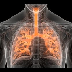
In the wake of the Covid-19 pandemic, medical imaging methods are playing a more prominent role, especially in relation to the primary manifestation of pulmonary disease and the tissue distribution of the angiotensin-converting-enzyme 2 (ACE 2) receptor. Currently, there is no consensus view to guide clinical decisions regarding imaging procedures such as radiography, computer tomography (CT), positron emission tomography (PET), and magnetic resonance imaging and the risk of exposing staff to infection.
This is based on findings from an article “A comprehensive review of imaging findings in COVID-19 - status in early 2021” published in the European Journal of Nuclear Medicine and Molecular Imaging.
The aim of the study was to depict the broader utility of medical imaging for detecting COVID-19-associated pathologies involving the nervous system and sensory, musculoskeletal, and cardiovascular systems, as well as renal, gastroenterological, and dermatological involvement.
One of the reasons for resorting to imaging procedures is that current RT-PCR methods for positive diagnosis is insensitive. CT is more sensitive than genetic testing for those patients already hospitalized. However, positive findings of ground glass opacities depend on the stage of the disease. Although there is very little reporting on PET/CT with [18F]-FDG in COVID-19, the available results correspond to earlier reports on viral pneumonias. Also, there is a high incidence of gastrointestinal involvement and cerebral findings in Covid-19.
Due to advances in Artificial intelligence (AI) and deep learning (DL), large datasets and powerful hardware based on graphics processing units, medical imaging has evolved significantly. During the pandemic, AI has helped in assisting with the diagnosis, stratification, prognosis and treatment of COVID-19 patients, for example the analysis of thoracic images and radiomic analysis of chest X-ray (CXR) images. The performance of AI systems in the discriminative diagnosis of COVID-19 is comparable to that of human practitioners.
At the moment, there is no indication for the use of PET/CT in routing clinical diagnosis or managing Covid-19. However, multi-modal imaging may shed light on pathophysiological processes involved in this new disease. For instance, well-established nuclear medicine research tools – like radiolabelling of immune or inflammatory cells for PET imaging – show how molecular imaging could lead to developing innovative therapies.
The authors acknowledged that the evidence gathered from across the field of medical imaging is limited to late 2020 and early 2021, thus knowledge pertaining to imaging results for mid- and long-term effects is lacking.
Source: European Journal of Nuclear Medicine and Molecular Imaging
Image credit: iStock
Reference:
Afshar-Oromieh, A et al. A (2021) Comprehensive review of imaging findings in COVID-19 - status in early 2021. Eur J Nucl Med Mol Imaging. doi:10.1007/s00259-021-05375-3


