

Ultrasound might not seem an obvious weapon against COVID-19, the disease caused by the new coronavirus, and its ensuing complications. Best known for helping doctors and expectant parents to view babies in utero, ultrasound sees inside the body by emitting and recording the echoes of high-frequency soundwaves. It’s similar to how bats navigate at night.
Ultrasound machines have become indispensable in hospitals since their debut in the 1950s, but by applying artificial intelligence to interpret those echoes, typically displayed as grayscale images, doctors are getting a new level of insight. And that’s helping them provide care for patients with COVID-19.
Ordinary ultrasound machines have become useful tools because they can help doctors monitor COVID-19’s tell-tale lesions just below the pleura, the membranes that envelop the lungs, and the body wall. “It has been underestimated, the power of ultrasound in the intensive care unit,” says Antonio Spera, the general manager for GE Healthcare in Italy who is leading efforts to ensure the country’s hardest-hit hospitals have critical equipment. “It’s now exploding.”
AI adds diagnostic speed. The set algorithms, which had been trained on hundreds of thousands of ultrasound images, can automatically detect irregularities and giveaway patterns in the grayscale and highlight them for clinicians. It can help doctors get a handle on the extent of an infection in a few seconds, allowing them to quickly make triaging decisions. The larger and more frequent the lesions, the more severe the case. AI that zeroes in on B-lines (see image below) and counts them has saved Chinese physicians valuable time, says Dr. Yangong Chao, director of critical care medicine at the First Affiliated Hospital of Tsinghua University in Beijing.
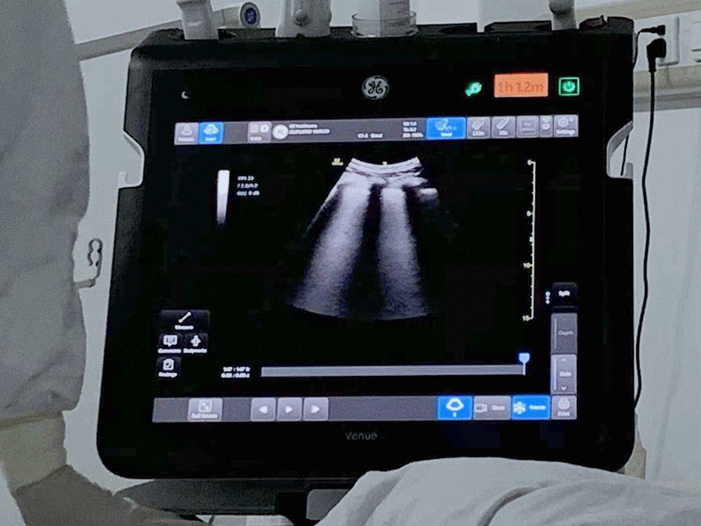
Top and above: The algorithms, which have been trained on hundreds of thousands of ultrasound images, can automatically detect subtle irregularities and giveaway patterns in the grayscale and highlight them for clinicians. Top and above images credit: GE Healthcare.
Mathias Goyen, chief medical officer for GE Healthcare in Europe, explains that ultrasound also has an advantage over computed tomography (CT) scans, the standard procedure to assess pneumonia in “ordinary” times. “In an ideal setting, CT is the imaging method of choice for COVID-19. But we don’t have an ideal setting — we’re dealing with an overflow of patients as part of a pandemic. In this COVID-19 crisis that is unfolding, ultrasound is often the imaging modality that is most readily available, compared to CT, at point of care.” Goyen added that “ultrasound could be the imaging method of choice for many patients on the ICU in this COVID-19 overflow situation.”
Researchers — who used lung ultrasonography to monitor more than a dozen patients in China with critical COVID-19 infections — have suggested ultrasound could be the imaging method of choice for many patients in an ICU as hospitals get busier. “Based upon our experience, we consider that lung ultrasonography has major utility for management of COVID-19 with respiratory involvement due to its repeatability, absence of ionizing radiation and point of care use,” wrote researchers from the critical care department at Xiangya Hospital in Changsha.
AI-enabled ultrasound also can help doctors get a handle on potential complications from the disease. For example, during the 1918 influenza pandemic commonly called the Spanish flu, some of the highest risk came from bacterial infections and adverse immunological reactions that were triggered by the virus, rather than from the virus itself, according to the U.S. Centers for Disease Control and Prevention.
Sepsis is the ever-lurking threat in such cases. It occurs when an immune system overreacts to the viral infection, releasing proteins called cytokines. A cytokine “storm” dilates blood vessels, making them leaky. That, in turn, can starve vital organs of oxygen, causing them to fail. Sepsis contributes to around 20% of all deaths worldwide every year, according to a recent study published by the journal Lancet. Emergency doctors can administer antibiotics, intravenous fluids and high-flow oxygen to treat sepsis, but early detection is critical for improving patient outcomes. That is where AI-assisted ultrasound is a doctor's ally.
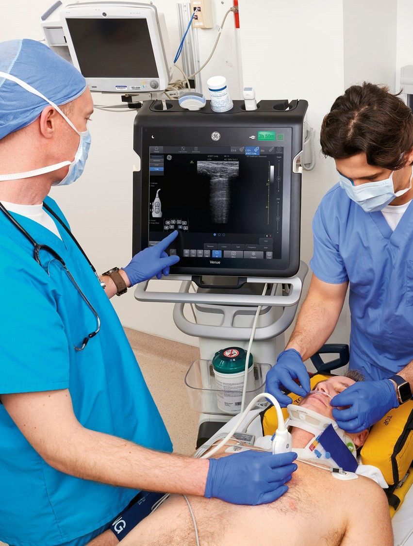
"In this COVID-19 crisis that is unfolding, ultrasound is often the imaging modality that is most readily available, compared to CT, at point of care,” said GE Healthcare's Mathias Goyen. The image shows an illustration of an ultrasound lung exam, not an actual COVID-19 situation. Image credit: GE Healthcare.
For example, doctors usually determine whether the heart is pumping enough blood through the primary arteries to end organs by taking an ultrasound measurement called Velocity Time Integral (VTI). Hardware with AI-enabled features, such as Venue and Venue Go, developed by GE Healthcare, can get that VTI reading in seconds, not minutes. That’s because once physicians have an image of the patient’s beating heart, they can tap a button on the screen (Auto VTI) and the system will automatically place a box where the organ’s left ventricle meets the aorta, the body’s main artery. (The sample box looks similar to the frame that pops up over people’s faces when you take a photo on a smartphone.)
Algorithms do the rest. Within seconds, they can analyze the outlined waveform showing the readings for the rate of blood flow in meters per second, and its volume in liters per minute. The technology dramatically reduces the number of keystrokes and the time it would take to calculate the flow rate manually. “For COVID-19 patients, the medical staff has to wear very heavy clothes, a face mask and multiple gloves,” says Chao with the First Affiliated Hospital of Tsinghua University. “So, when we use these automatic functions, it not only saves time but a lot of energy in not having to do all these steps. It’s very useful.”
The machine can show VTI trends to highlight changes in response to therapy. That allows medical workers to regulate the fluids supplied to the patient.
Ultrasound machines help providers monitor COVID-19 patients in other ways. Their portable, compact and ergonomic design — compared with, say, computed tomography scanners — means they can easily adapt to various clinical spaces, such as emergency rooms, ICUs and operating rooms, where some of the most serious COVID-19 cases end up. The GE Healthcare Venue and Venue Go machines also sport a seamless flat display without knobs, keyboards or creviced surfaces. The display is easy to wipe down, which helps reduce risk of infections. “I have more confidence that I can clean the device better than other devices because there are less nooks and crannies that you need to clean,” says Dr. Bilal Jalil, a pulmonologist and critical care physician at Baylor Scott and White Health in Dallas.
That’s crucial, because the world’s healthcare workers are on the front line against COVID-19, and they will be fighting the good fight daily in the coming months.