Three-dimensional (3D) printing is increasingly being used in medical practice, but its implications for healthcare may be complex, according to a commentary published in JAMA. Although questions remain about how best to use the technology, experts say that 3D printing is already changing patient care. In fact, 3D printing has been used in dentistry for more than 10 years, they note, because it enables the creation of moulds for many dental implants.
"While not a panacea, 3D printing is increasingly finding its place in patient care, from its expanding use in surgical planning to the vision of printing whole new organs for transplantation," writes Mark H. Michalski, MD, from the Investigative Medicine Program, and Joseph S. Ross, MD, from the Robert Wood Johnson Foundation Clinical Scholars Program, both at Yale University School of Medicine in New Haven, CT.
"Indeed, 3D printing may serve as a means of distributing manufacturing in the same way that the Internet distributes information," the authors point out.
Now the technology helps head and neck surgeons to make preoperative models for complex surgeries. "For example, several facial reconstructive surgeries are performed by first harvesting the fibula, which is then fashioned in the operating room into new bony structures," the Yale authors say. "These surgeries can now be augmented using computer planning programs to generate surgical plans that determine the ideal way to harvest and incise the fibula to create a reconstructive graft."
In addition, researchers outside hospitals and clinics are using 3D printing to create medical devices for a wide range of conditions that are suited to an individual patient's anatomy. The authors write that a biocompatible polymer splint has been used to prevent airway collapse in one case of neonatal bronchomalacia. The splint was designed to be naturally resorbed within three years.
Safety and Regulatory Issues
3D printing also has been utilised to make prosthetics, with digital blueprints made freely available for download and reproduction, according to the JAMA article. However, few studies have assessed how 3D printing has performed regarding safety and regulatory issues. "At this early phase, it is unclear what the ultimate value of 3D printing will be for health and how it will specifically affect outcomes," the authors note.
Questions also remain as to how regulation may affect use of 3D printing. "As 3D printing continues to integrate into medical practice, physicians and patients face the challenge of understanding this complex technology, taking advantage of its potential, and weighing its potential risks," they conclude.
"I think it's very prescient for [JAMA] to publish this," Leonard S. Marks, MD, a urologist at the University of California, Los Angeles, Medical Center, told Medscape Medical News. He co-authored a study published earlier this year on using 3D imaging to create moulds for prostate organs after their removal.
"There are lots of applications in medicine for 3D printing," Dr. Marks said. "Our interest in this began about three or four years ago. We are interested in the correlation between prostate cancer seen on [a magnetic resonance imaging] image and prostate cancer as it appears in the organ after it's removed."
3D printing helps provide information that is used to determine treatment levels, Dr. Marks added.
"It's allowed us to predict what's actually in the prostate. We've never had that opportunity before," the doctor explained. "It allows us to find the serious cancers more quickly and to find the insignificant cancers, which are very common in the prostate, and put them in proper perspective more quickly and to save people the worry that they may have a serious disease when in actuality there's not. You know who's got a bad one and who doesn't."
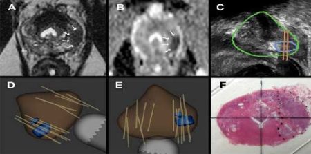
Top Image
Legend:
A. T2-weighted axial MRI demonstrating a lesion in the left peripheral prostate.
B. Diffusion weighted MRI showing restricted diffusion (ADC value of 562) within the lesion.
C. Real-time ultrasound image of the lesion (outlined in blue) deriving from MRI fusion in Artemis
device.
D. and E. 3D reconstruction of prostate, based on ultrasound scan, showing lesion from MRI fusion (in blue) within the model, (D) saggital and (E) transverse views. Tan lines, which are image-captured biopsy sites, show sites of both systematic and targeted biopsy cores. Targeted biopsies in this patient revealed Gleason 7 prostate cancer.
F. Radical prostatectomy specimen showing tumour (dotted line) in whole mount section. Histologically, tumour was a 2cm Gleason 7 cancer in the left peripheral zone.
Source: Medscape.com
Image Credit: UCLA
Latest Articles
MRI, Surgery, prostate cancer, JAMA, 3D printing, dentistry
Three-dimensional (3D) printing is increasingly being used in medical practice, but its implications for healthcare may be complex, according to a commenta...


