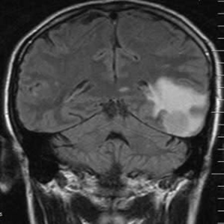
T2-weighted magnetic resonance imaging (MRI) hyperintensity assessed visually in the corticospinal tract (CST) lacks sensitivity for a diagnosis of amyotrophic lateral sclerosis (ALS). In a new study, researchers sought to explore a quantitative approach to fluid-attenuated inversion recovery (FLAIR) MRI intensity across a range of ALS phenotypes. The study findings show that FLAIR MRI encodes quantifiable information of potential diagnostic, stratification, and monitoring value.
ALS, a neurodegenerative disorder, is predominantly a clinical diagnosis with significant heterogeneity in rate of disability accumulation. Therapeutic trials rely on survival or change in disability accumulation rate as the primary endpoints. Biomarkers are therefore a research priority.
T2-weighted hyperintensity of the CST was the first documented abnormality in standard clinical MRI sequences in ALS, but proved insufficiently sensitive based on only visual inspection. Recent studies though have begun to revisit the diagnostic potential of T2-weighted sequences in ALS. In the current study, researchers hypothesised that a quantitative approach to assessing FLAIR intensity has diagnostic and stratification biomarker potential in ALS. Their aim was therefore to apply this technique to the principal motor tracts of the brain in ALS patients across a range of clinical phenotypes and a group of healthy controls.
The study included 33 classical ALS patients – 10 with a flail arm presentation and six with primary lateral sclerosis (PLS) – who underwent MRI at 3 Tesla. Comparisons of quantitative FLAIR intensity in the CST and corpus callosum were made between 21 healthy controls and within patient phenotypic subgroups, some of whom were studied longitudinally.
The results showed that mean FLAIR intensity was greater in patient groups. The cerebral peduncle intensity provided the strongest subgroup classification. In addition, the researchers observed that FLAIR intensity increased longitudinally. The rate of change of FLAIR within CST correlated with rate of decline in executive function and ALS functional rating score.
"The small subgroup of PLS patients demonstrated some of the highest FLAIR intensities, but there was no simple relationship with burden of UMN [upper motor neuron] signs," the authors note.
The finding of highest FLAIR intensities in PLS patients is building on other neuroimaging research findings in this subgroup. This highlights the need for earlier diagnosis in PLS patients to improve the feasibility of earlier therapeutic intervention as well as care planning, the research team points out.
The study has several limitations, including the relatively small numbers of participants in the flail arm and PLS subgroups, as well as the longitudinal cohort.
The team says a more comprehensive cognitive (and behavioural) assessment, and direct comparison with other MRI markers of white matter pathology, for example, Apparent Diffusion Coefficient (ADC) and diffusion tensor imaging (DTI), would be desirable in future studies.
Source: Academic Radiology
Image Credit: Nevit Dilmen
Latest Articles
FLAIR MRI, amyotrophic lateral sclerosis, fluid-attenuated inversion recovery
T2-weighted magnetic resonance imaging (MRI) hyperintensity assessed visually in the corticospinal tract (CST) lacks sensitivity for a diagnosis of amyotrophic lateral sclerosis (ALS). In a new study, researchers sought to explore a quantitative approach


