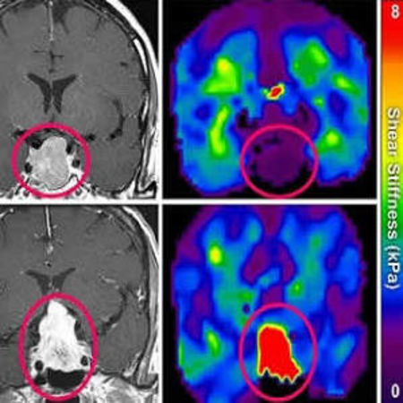
Researchers at Mayo Clinic have used magnetic resonance elastography (MRE) to assess pituitary tumour stiffness by measuring waves transmitted through the skull into pituitary macroadenomas. MRE, often called “palpating by imaging”, non-invasively identified tumours removable by simple versus complex surgeries, according to a study published in the journal Pituitary.
MRE captures snapshots of shear waves that move through the tissue and create elastograms, or images that show tissue stiffness. Ninety percent of pituitary macroadenomas (PMAs) are soft — nearly the consistency of toothpaste. Using MRE, hard PMAs can be identified and the more extensive craniotomy can be planned before starting the surgery, which makes the more invasive procedure less taxing for both the surgeon and patient.
In the new study, the Mayo team performed pre-surgical MRE examination of the PMAs of 10 patients. The MRE measurements were compared to tumour classifications made by inspection of the tumour during surgery. The surgeons categorised six tumours as soft and four tumours as medium. No tumours were deemed to be hard. The comparison of the MRE results and reports of stiffness by the surgeons when the tumour was removed and inspected were in close agreement, which was confirmed by statistical analysis.
Although brain MRE is not yet widely available, the technique is now routinely used by surgeons at the Mayo Clinic to plan the best procedure for the removal of PMAs as well as several other types of brain tumour. An important aspect of some of the other brain tumour types, which the Mayo surgeons are finding extremely useful, is the ability of MRE to identify tumour adhesion to the brain, says John Huston III, MD, professor of radiology at the Mayo Clinic in Rochester, MN, and senior author of the study.
Adhesion refers to whether the brain tumour and healthy brain tissue are connected by an extensive network of blood vessels and connective tissue. This is in comparison with a tumour that is in the brain but is isolated from healthy tissue. When MRE is used to analyse this aspect of the tumour, it clearly identifies those that are non-adhered, showing a border around the tumour through which there are no vascular connections. In contrast, MRE of adhered tumours show no border between the tumour and healthy brain, indicating extensive vascular and soft tissue connections between brain and tumour.
According to Prof. Huston, his team is continuing to apply MRE to diagnosis of other brain disorders. In addition to characterising focal brain disorders such as tumours, the team is testing the potential for MRE to provide diagnostic information about diffuse brain disease. MRE brain stiffness patterns are also used to identify different types of neural disorders including dementia.
Source & Image: National Institute of Biomedical Imaging and Bioengineering
Latest Articles
magnetic resonance elastography, MRE, tumour stiffness
Researchers at Mayo Clinic have used magnetic resonance elastography (MRE) to assess pituitary tumour stiffness by measuring waves transmitted through the skull into pituitary macroadenomas. MRE non-invasively identified tumours removable by simple versus


