HealthManagement, Volume 16 - Issue 2, 2016
‘Making It Work’ vs. ‘Making It Fit’

When it comes to design and construction, understanding a hospital’s ongoing needs is “the single most essential piece”. That is why siting imaging equipment often gets left to the last consideration, because the technical criteria have more objective answers. So, although ‘making it fit’ is rarely a problem, the more important goal will always be ‘making it work, well’.
US Guidelines on MRI Suite Design and Safety
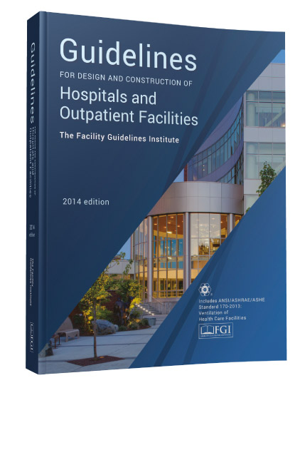
The most frequently used design standard in the US is the Facilities Guidelines Institute (FGI) “Guidelines” publication that covers all aspects of hospital design (not just imaging), and is updated every four years. I had been a subject matter expert on the imaging sections for the last two editions (2010 and 2014), and I am currently serving as a full committee member working on what will be the 2018 edition.
Imaging, in general, has been a challenge for FGI as the technology has been moving so quickly, particularly when compared with a four-year process for re-releases of the ‘Guidelines’ books. The perfect examples of these are the two comparatively new ‘hybrids’ – hybrid modalities (e.g. PET/CT, PET/MR) and hybrid operating rooms (ORs).
Hybrid ORs (pairing imaging with surgery) aren’t an entirely new concept, but the depth and breadth of image-enabled procedures has grown substantially in the last ten years. If the line that separated intervention from surgery was blurry before, it is almost obliterated now. Today, it is impractical to define interventional rooms by a room name (‘cardiac catheterisation lab’, ‘interventional radiology room’, etc.), without defining what types of procedures might occur within. The level of intervention now capable in an ‘interventional suite’ is really akin to what would have been done exclusively in a surgical theatre ten years ago, making the prospective discussion of what types of procedures will be performed, and how the hospital will handle complications, essential initial planning tools.
To this end, the 2018 ‘Guidelines’ publication is planning to introduce an acuity/intervention classification system. Imaging rooms will be designated ‘Class One’, ‘Class Two’, or ‘Class Three’ depending on the clinical utilisation of the room. Basic diagnostic (only) imaging functions will be ‘Class One’. Image guided minimally-invasive procedures or imaging requiring respiratory support will be ‘Class Two’. And open surgical environments with imaging will be ‘Class Three’. Each classification will have distinct requirements for clinical support, infection control, and sterility.
Hybrid imaging modalities are similarly complicated, though for different reasons. The clinical process and workflow for, say, a PET patient, and a CT patient, are profoundly different. When these two modalities are fused into a single piece of imaging equipment (PET/CT), the least of the designer’s problems is the room in which the scanner goes. Yes, the room likely needs to be on the order of 1.5m longer than a conventional CT scanner room, but that’s comparatively easy.
PET patients need to have injections of radioisotopes, and typically need to remain still for 45 minutes to an hour following injection and prior to imaging. Whereas CT is today a speedy exam (a cardiac CT examination can often be accomplished in less time than it takes to get the patient comfortably positioned in the correct spot on the table), PET exams have a much different timetable.
And - in the US, at least - CT radiographers and PET radiographers have entirely different training and certifications. Even the departmental structures (radiology vs. nuclear medicine) are most frequently separated. So, staffing and training for hybrid modalities can become a challenge, along with questions of authority and ‘ownership’ of a hybrid. This aspect will only get more complex with new hybrid MRI linear accelerators that are currently available, and future three-way hybrids.
In both ‘hybrids’ (hybrid OR and hybrid modalities), the circumstances really demand that facility planners and designers have very detailed conversations with the hospital clients to understand their operational wants and needs prior to designing the spaces. While we do give these spaces labels that might suggest that they are off-the-rack items to be selected, they really need to be approached with the expectation that they are really bespoke, tailored to the specific needs of the client.
Universality of Guidelines and “Cross Border” Compatibility Issues
In the US , design and licensure standards are set by each state, individually. Fortunately, the overwhelming majority of them recognise the FGI ‘Guidelines’ for their state standard. Even those that have developed their own standards often base their criteria (largely) on the FGI ‘Guidelines'. National accrediting bodies, such as The Joint Commission refer to FGI as the default standard, if a facility is in a location that doesn’t have a state or local healthcare design standard.
‘Guidelines’ is not quite universal in the US , but it is far and away the single most widely recognized design standard for hospitals and healthcare construction.
With the new classification system, it means that hospitals and designers will need to be having more in-depth predesign conversations before MRI projects begin. ‘Will this suite support high-acuity patients? Will it support heavy sedation / general anaesthesia patients? Will it support image-guided procedures?’ The answers to these questions will help drive the design criteria for the imaging suites.
If imaging equipment manufacturer ‘typical’ MRI suite templates were ever sufficient (and I would argue that they never have been), they certainly won’t be once the acuity classification system is introduced. The same will be true for other imaging modalities.
Keeping Up With Regulations – Another “Magnetic Elephant in the Room”?
There absolutely are some design expectations that are being driven by newer technology and clinical applications, but from an MRI safety point of view, what designers are being asked to do, today, is really what was outlined almost 15 years ago in the original American College of Radiology (ACR) White Paper on MRI Safety (subsequently rewritten and reissued in 2004, 2007, and most recently in 2013 as the ACR Guidance Document on MR Safe Practices). What is being codified as design standards today has really existed as an industry best-practice for well over a decade. It was unfortunate that architects, engineers, equipment planners and facility managers didn’t uniformly embrace MRI safety design best practices, but the new design standards will help to assure that newly renovated and constructed MRI suites will adhere to these as minimums.
The Future of Design in Radiology
Radiology today is being compressed. It used to be that providers offered conventional X-ray, ultrasound, or bone densitometry as necessary services, but counted on CT and MRI to generate the overwhelming bulk of their revenue / profit. In the US , the reimbursement levels from insurers were high enough for CT and MRI that it really didn’t matter if the other services paid for themselves. In fact, some pretty poor design and operational practices were institutionalised in the US when these services were awash in reimbursement. Today, with decreasing reimbursement, outcome-based payments, and resulting pressures on increasing patient volume, many existing facilities / providers in the US are finding themselves ill-equipped to respond to the new paradigm.
Radiology facilities today need to focus on operational efficiency as never before. If a CT study can be complete in less than five minutes, then that CT might be able to image 6-8 patients per hour. If the facility design is a carbon copy of prior imaging suites from years ago, it likely doesn’t have the patient management / preparation spaces and features that are necessary to support the capacity potential of the scanner. Why spend 1-2 million Euros on a high throughput scanner, only to cut that potential in half through a poorly planned facility?
The difficult truth to this is that there are no copy-and paste prototypes for the new economic realities for medical imaging. Architects and equipment planners who simply provide a ‘garage’ for the piece of equipment, and don’t help the hospital crystallise their operational goals, are doing their clients a grave disservice.
Hardware Fatigue, Used MRI Systems and Software Updates
Medical imaging equipment just continues to get better and better… much of it lasting longer and longer. This has been a benefit for hospitals riding out the economic downturn of 2008, as there wasn’t the capital available to replace equipment on the prior schedule. Older equipment and used equipment isn’t necessarily bad, and it can reduce first costs (though I argue that operational efficiencies should always be a higher priority than first costs), but it does necessarily limit the features and functions that are available to the hospital.
As an example, I have a first generation iPad. It still works… there is nothing physically wrong with it. But it cannot accept the latest system software updates, which means that it can’t accept new programmes that depend on the newer system software. I can still check my email, browse the web, or download music, but I can’t take advantage of applications that have been released in the last few years. Older CTs and MRI s are quite similar in this respect… they may work equally well as when they were first released, but they will likely never compete with newer equipment in terms of the range of functionality that they can support.
Physical Screening and Changing Areas
Screening and changing areas are also essential parts of this emphasis on efficiency and throughput, along with patient sub-waiting areas. These are also elements that are not typically portrayed in manufacturers’ equipment siting prototypes, and therefore don’t get appropriate consideration when designs begin with vendor templates.
When working with clients to design imaging spaces, I like to start with their ultimate end-goals (e.g. ‘x number of imaging studies / day or week’), and back up from there. When the goal is ‘site the MRI scanner we’re purchasing’, that really only addresses the short-term goal, but not the persistent condition that the hospital will need to live with for years and years. I prefer to identify that ongoing condition, and the hospital’s expectations for that. To a skilled radiology designer, simply siting MRI equipment is not difficult. The challenge is in siting it such that it brings the greatest value to the hospital.
Access Control
Ionising radiation-emitting modalities, like X-ray and CT, have one set of controls that are typically tied to the operation of the X-ray tube. MRI and nuclear medicine have different access controls (both from CT and from each other) based on persistent risks. Each of these need to be honored, individually. Particularly when we begin mashing together modalities with very different access restrictions into hybrids (i.e. PET / MRI ), the overlapping and conflicting access controls can get complicated. It often takes some creativity and a very careful evaluation of the operational model (for example, is it the hospital’s intention to do a large proportion of their studies as simultaneous PET and MRI exams, or will the majority of the patients be only one or the other?) to develop a layout that honours the safety and access restrictions of each of the combined modalities, but also allows for efficient operation.
Accommodation
of Site-Specific Clinical and Operational Requirements
This is the single most essential piece. It is, after all, the part about understanding the hospital’s ongoing needs. When it comes to siting imaging equipment, this often gets left to the last consideration, because the technical criteria have more objective answers. I understand this sequence for comparatively inexperienced designers, but in my opinion it is totally backwards.
‘Making it fit’ is rarely a problem. The more important goal will always be ‘making it work, well’. For this, I never start with the equipment manufacturer’s minimum technical siting criteria, but with the owner’s clinical and operational requirements… both today’s and a reasonable forward-looking notion of what might be added in the years ahead.
Key Points
- The 2018 facilities’ guidelines will see imaging rooms designated
depending on the clinical utilisation.
- Staffing and training for hybrid modalities can become a
challenge, along with questions of authority and ‘ownership’.
- There are no copy-and-paste prototypes for the new economic
realities for medical imaging.
- Architects and equipment planners who simply provide a
‘garage’ for the piece of equipment are doing their clients a grave disservice.
- To a skilled radiology designer, simply siting MRI equipment is not difficult. The challenge is to bring the greatest value to the hospital.
References:
ACR (2004) MR Safety. White Paper. [Accessed: 17 June 2016] Available from http://www.acr.org/quality-safety/radiology-safety/mr-safety
ACR (2007) MR Safety. White Paper. [Accessed: 17 June 2016] Available from http://www.acr.org/quality-safety/ radiology-safety/mr-safety
ACR (2013) Guidance Document on MR Safe Practices. [Accessed: 17 June 2016] Available from http://www.acr.org/quality-safety/radiology-safety/mr-safety
Facilities Guidelines Institute (FGI) (2010) Guidelines for Design and Construction of Health Care Facilities. [Accessed: 17 June 2016] Available from http://www.fgiguidelines.org
Facilities Guidelines Institute (FGI) (2014) Hospital/Outpatient Guidelines. [Accessed: 17 June 2016] Available from http://www.fgiguidelines.org
Facilities Guidelines Institute (FGI) (2014) Residential Guidelines. [Accessed: 17 June 2016] Available from http://www.fgiguidelines.org