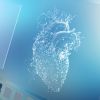


A team of scientists at the Universidad de Valladolid and the Centro Nacional de Investigaciones Cardiovasculares (CNIC) has developed a new method for identifying 3D features of scar tissue formed after a myocardial infarction.
The new method allows 3D transmural mapping of scar tissue in the infarcted muscle which enables detailed characterisation of the morphology of the damaged tissue and provides an accurate measure of infarct size relative to myocardial wall thickness.It uses conventional 2D delayed gadolinium-enhanced CMR sequences and requires only a limited number of slices.
Image Credit: CNIC
According to the team, the method is compatible with standard cardiac magnetic resonance (CMR) sequences and can shorten the time needed for image acquisition, thus easing access to nuclear magnetic resonance scanners which are already quite in demand.
The authors state that this novel method could prove to be an efficient approach in clinical practice after manual or automatic segmentation of myocardial borders. Their findings show that low scar transmurality on CMR (below 10% of ventricular wall thickness for 3D sequences or 20% for 2D sequences) is associated with ventricular tachycardia. In addition, results show that patients with low scar transmurality values had a higher probability of ventricular tachycardia recurrences during long-term follow-up.
Source: CNIC